 |
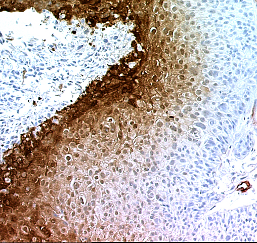
| Immunoperoxidase staining for hypoxia (Hypoxyprobe-1 adducts; brown stain) and blood vessels (Factor VIII, red stain) in a formalin fixed, paraffin embedded tissue section from a human squamous cell carcinoma of the uterine cervix. The cancer patient had been infused with a dose of 0.5 grams per meter2 of Hypoxyprobe™-1 and a biopsy of the tumor taken 24 hours later. Note the gradient of increasing immunostaining for hypoxic cells across the section from the blood vessel on the right to necrosis on the left of the image (magnification x100). Multiple images such as these have been used to quantify the extent of hypoxia that ranges from 0 to 20% for cervix squamous cell carcinomas. |